In literature, the human heart is often associated with valor and invincibility. From an anatomical perspective, this image is slightly inaccurate since cardiomyocytes — cardiac muscle cells — are post-mitotic and stop dividing shortly after one's birth.
On Tuesday, April 7, the Department of Chemistry welcomed Herdeline Ann "Digs" Ardoña from the University of California, Irvine, for a seminar titled "Precision engineering of macromolecular systems as transducer biomaterials." During this seminar, Ardoña discussed how her group is attempting to change the narrative around cardiac muscle cells by developing organic materials that promote cardiomyocyte maturation. This can potentially allow for the design of artificial cardiac tissue environments capable of regeneration.
Ardoña's research revolves around transducer biomaterials. “These are materials that allow for the conversion of cues, such as optical, electronic, chemical and mechanical phenomena, at the interface of electroactive or electrogenic materials,” she said. From a therapeutic standpoint, such materials may be applicable in tackling conditions exacerbated by the post-mitotic nature of cardiomyocytes, like heart attacks.
Currently, cardiomyocytes can be derived from stem cells, undifferentiated cells that can develop into many different cell types. However, the cardiomyocytes produced by such protocols are immature.
“When I say immature, its electrophysiology doesn’t match the adult [cell’s] physiology, and that’s mainly because the structural features of these cells [as you can see], are very different in terms of [its] complexity,” Ardoña explained.
In the search for prospective alternatives, Ardoña says she is motivated by a central question. “Can we provoke the control of cardiac structure and function through cues delivered to artificial muscle environments?” she asks.
Ardoña then delved into the design criteria her group follows while mimicking extracellular environments. First, the bioengineered cells must support some form of intercellular communication, which is carried out using ion channels that open and close in response to chemical and mechanical signals in biological settings. A soft environment must also be created to ensure the engineered material is biomimetic — meaning it matches the appearance and feel of natural muscle cells. Finally, local tissue alignment is especially critical for cardiomyocytes. This refers to the extent to which muscle cells interact with the extracellular matrix, the network of proteins that surrounds them, which affects their contractile properties, like the force they produce and the velocity with which they constrict and relax.
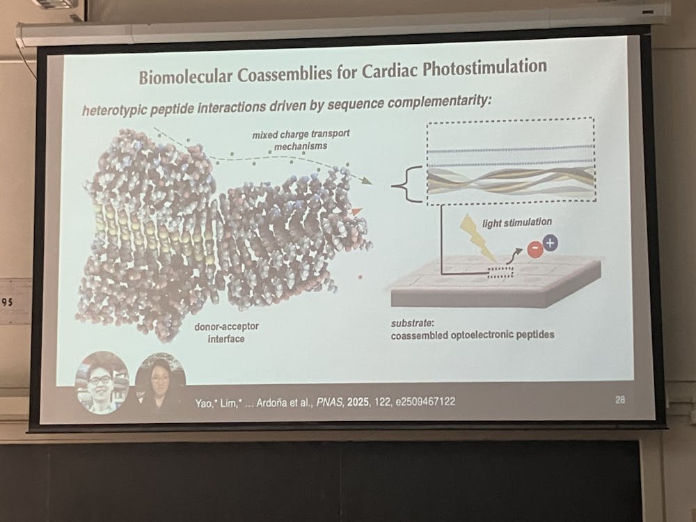
Her group has already taken steps toward this goal. For instance, they've developed a method to micropattern a photosensitive polymer onto cardiac muscle cells. This enables the generation of microcurrents that stimulate the cells. Using a short clip of a setup where the beating of cardiomyocytes caused a cantilever to oscillate, Ardoña demonstrated this polymer at work.
“We would pulse one hertz of light, and we would see that the cantilever is really interestingly following the pulse of light, and then when we would pulse two hertz of light, we would be able to find the pacing [is] much faster,” she described.
Ardoña acknowledged that these solutions are still limited at the moment. One weakness is that the matrices used to grow artificial tissue are water-insoluble, requiring strong solvents to process the layers.
”We want to move towards more biomimetic or bio-inspired sources,” Ardoña suggested.
It is essential to remember that this project does not exist in isolation; rather, it builds on Ardoña's lab's work in peptide coassembly. Based on charge, peptide assembly can be ordered or disordered. Cardiomyocyte growth requires ordered environments to conduct electrical signals efficiently. A disordered environment could impede signal transfer, like how improper wiring leads to a faulty circuit.
“There are other designs that we are exploring, and I would say that in terms of the future we’re modulating the current stimulation pathway, but we also want to impart specificity in how they [transducer biomaterials] are delivering [a specific cue],” Ardoña explained as she reflected on what the future holds.
She is hopeful that she can use the systems she is pioneering to stimulate the maturation of excitable cells other than cardiomyocytes. In this way, she wishes to push the boundaries of tissue engineering, paving the way for a future in which cardiomyocytes matured using transducer biomaterials can replace dead tissue after a heart attack. Perhaps we can make the human heart invincible after all.


