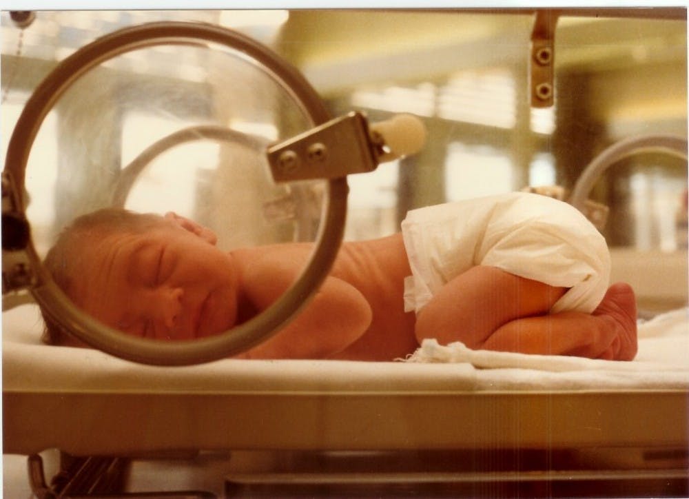
According to the World Health Organization (WHO), approximately 15 million babies are born preterm each year. Nearly a million babies die due to preterm birth complications, making preterm birth a leading cause of child death. On top of that, survivors of preterm births often develop learning, visual or auditory disabilities.
Preterm birth, also referred to as premature birth, is when babies are born prior to 37 weeks of pregnancy. It can be divided into more detailed sub-categories based on gestational age.
A variety of factors can cause preterm birth, mainly represented by spontaneous preterm birth that is currently tough to predict or prevent. Preterm birth is known to be often preceded by preterm premature rupture of the membranes (PPROM). In addition, most cases of preterm birth are termed “idiopathic,” meaning that there is no identifiable cause, such as infection or bleeding. Therefore, it remains a crucial task to find biological mechanisms that are responsible for idiopathic preterm births.
Recently, in a study published by the American Association for the Advancement of Science (AAAS), a group of scientists lead by Lydia L. Shook of the Yale University School of Medicine shed light on a potential cause for idiopathic preterm birth by examining the formation and deposition of calciprotein particles in more than 100 pregnant women.
Shook’s research group hypothesized that “calcium and phosphate-rich amniotic fluid (commonly known as a pregnant woman’s “water”) harbors potential for calciprotein particle formation and its aberrant deposition in the amniochorion (fetus membrane), resulting in the pathogenic effects involved in PPROM and spontaneous preterm birth.”
Calciprotein particles, a mineral-protein complex, are not only associated with diseases like arthritis and atherosclerosis, but these particles are also involved in abnormal calcifications that arise in three conditions: “a suitable extracellular matrix framework, excess inorganic phosphate and ionized calcium, and reduction or absence of mineralization inhibitors,” the study states.
The formation of calciprotein particles is caused by the imbalance between the production of hydroxyapatite (HA) and the endogenous mineralization inhibitor protein fetuin-A. Fetuin-A is an important protein that keeps minerals suspended in a solution. However, when the binding sites of fetuin-A are completely occupied, calciprotein particles precipitate out of the solution. These particles are then deposited in non-skeletal tissue, where they cause pathogenic effects.
Although the researchers did not discover any significant differences in phosphate or calcium concentration, they did find significant reduction in fetuin-A concentration from the amniotic fluid collected in idiopathic PPROM cases compared to the term control groups. This reduction is enough to disrupt the balance to favor the synthesis of calciprotein particles, further supporting the research group’s hypothesis that calciprotein particles play a role in PPROM.
To explain how the production of calciprotein particles can cause PPROM, the researchers experimented the effects of calciprotein particles on human amniochorion tissue cultures. Their results demonstrated that calciprotein particles lead to the breakdown of connective tissue, making them more prone to rupture.
The researchers proposed two possible explanations to this phenomenon. First, the formation of calciprotein particles “hijacks homeostatic amniotic fluid proteins and minerals that are necessary to maintain fetal membrane integrity.” Second, the formation of calciprotein particles “create actively toxic intermediates analogous to soluble protein oligomers formed in the process of protein misfolding.”
This study raises more attention on the role of calciprotein in birth disorder. It also suggests the importance of developing therapies that maintains the balance between HA and fetuin-A. The researchers state that future investigations are needed to understand what triggers the reduction of fetuin-A to have a better grasp about preterm birth.


