According to the World Health Organization, approximately 250,000 to 500,000 people suffer from spinal cord injuries each year. A lot of these injuries are due to preventable causes, such as car crashes and falls.
Spinal cord injuries often lead to mental disorders, with an estimated 20-30 percent of those affected showing symptoms of depression.
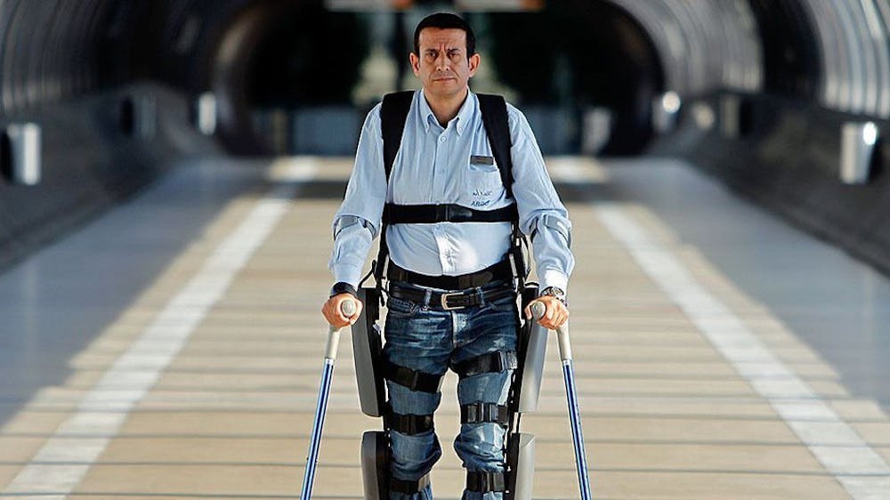
In addition, there still remain various obstacles that people with disabilities face in modern society. For example, children with spinal cord injuries are less likely to attend school, and adults with the injury face a higher rate of unemployment.
Achieving significant recovery from spinal cord injuries is a continual challenge. However, in a recent study done by Javier Ganz and colleagues from Tel Aviv University in Israel, the research team presented a promising stem cell treatment in mice.
They used human oral mucosa stem cells (hOMSC) embedded in scaffold matrix to help repair spinal cord injuries.
Past research has shown that hOMSCs can be induced to function like astrocytes that secrete factors aimed at enhancing neuroprotection, cell growth and differentiation.
Scaffolds are another crucial factor that provide an environment where cells can proliferate, attach and differentiate. In their experiment, the researchers created a biodegradable, porous scaffold out of equal parts of copolymers known as PLGS and PLLA.
“PLGA was selected to provide flexibility, whereas PLLA was chosen to provide stiffness,” Ganz said in a press release.
Ganz and colleagues hypothesized that combining hOMSCs with a PLGA/PLLA scaffold would create a device that helps rats recover from spinal cord injuries.
The researchers found that 42 percent of the rats treated with hOMSCs were able to support their hind limbs and showed improved walking abilities during the first three weeks.
Although the improvement peaked after five weeks, the effects were persistent enough to last until the experiment ended. The recovered mice progressively revealed similar walking patterns to those of normal mice.
To examine the extent of reconnection in the injured areas, the researchers used techniques such as MRI diffusion tensor imaging and motor evoked potentials (MEPs).
In their MRI data, there were no signs of reconnection three days after the injury across all groups. However, there was partial reconnection on the 56th day in the rats treated with hOMSCs.
Interestingly, the researchers found that mice treated with only the scaffold exhibited a lesser degree of recovery. Their results support other previous studies that demonstrated scaffolds alone can promote recovery.
The researchers also observed an on/off effect in the treated group, in which the mice were either responsive or unresponsive to the treatment. They believe that the implant position contributes to the distinct results.
“Since the minimum requirements for eliciting substantial recovery have yet to be defined, subtle differences in the scaffold and its position related to the spinal cord stumps may have selectively favored restoration of some but not all tracts,” the study said.
While the results in this article do not solve the problems of spinal cord injury and those associated with it, they do provide promising methods that urge the need to understand the mechanisms behind recovery.
According to the senior author of this study Shulamit Levenberg, even if there is still some way to go before the research can be applied in humans, the study’s findings are heading toward a promising route.


