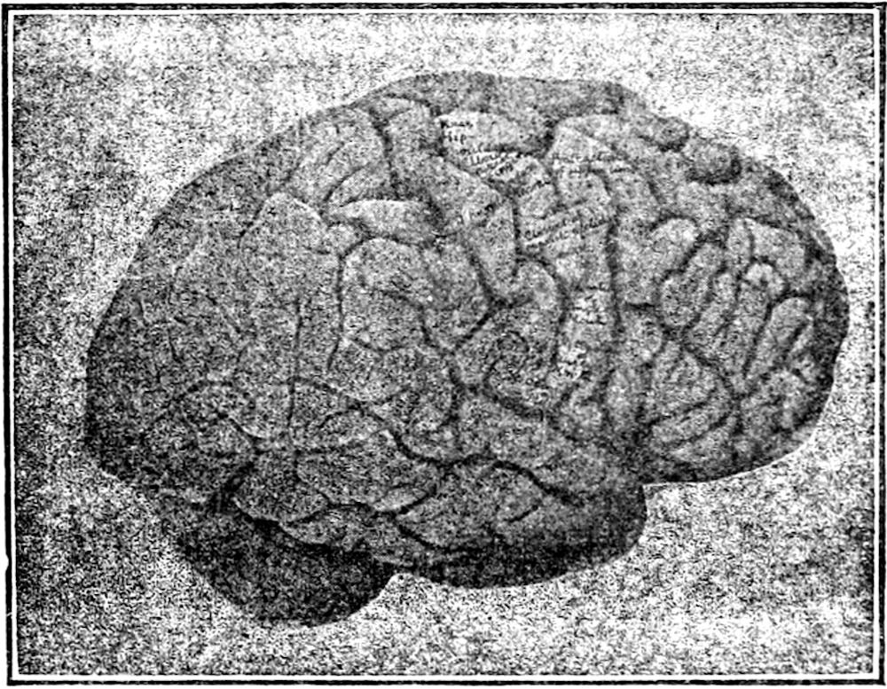
The diagnosis of psychiatric disorders can be a challenging task. The difficulties involved in standardising and quantifying measures of abnormality have been well known within the field. Having identified this struggle, Sridevi Sarma, professor for biomedical engineering at the Whiting School of Engineering, presented a seminar detailing her team’s work on a neural health index on Sept. 9.
Approaching the field of neuroscience from an engineering background, Sarma was able to identify key interdisciplinary connections that enabled her to think of the brain from a new perspective.
“What I've learned is that when you're trying to understand the complex system, in this case the networked brain, you actually don't need to understand all its details, its dynamics, in order to control it… If you think of a bicycle, I would argue it's somewhat of a complex system. If you talk about the laws of motion, the equations of motion that govern the bicycle dynamics… I'm confident that none of us are simulating these equations in our head to figure out how to control the bicycle,” Sarma explains.
The way to achieve this control, Sarma explains, is through understanding and synthesising the various sensory inputs involved, such as proprioception or vision. This enables the rider to create a model of the bicycle which they can then operate. A similar principle becomes transferrable for a practical model of the brain. Sarma’s lab works on several kinds of biomarkers and imaging data points in order to arrive at a simple, functional model of the human brain.
Beyond just a model, what distinguished Sarma’s approach was her focus on clinical applications. She argued for the limited capabilities of medical practitioners to regularly measure and monitor abnormalities in brain function — such practices are not integrated directly into most typical check-ups, and often disorders are only identified when it becomes too late to take action. Sarma’s team aimed to create a screening test that could successfully fill this gap.
“I want a number between, say, zero and one that says: zero, you don't have a healthy brain; one, you have a very good brain… it should distinguish a healthy brain from a not healthy brain… it should scale with the severity of the disease… it should modulate the treatment,” Sarma shared.
The concept that led to Sarma’s method involved the heavy engagement of a graduate student in her lab, whose varying field of expertise and willingness to challenge accepted ideas were identified by Sarma as key contributors to the success of the project. Across the board, collaboration played a crucial role: from working with other professors to cross-check mathematical calculations to drawing on the insights and data of active medical practitioners.
The central principle around which Sarma and her team were able to build their health index was entropy: the notion that there would be greater dispersion in a system while at rest and more concentration during focused activity. They noted this was visible across several biological systems. For example, schools of fish would be dispersed in normal conditions and gather together when avoiding predators. Similarly, brain activity shows no bias in location when at relative rest, but it becomes concentrated in specific functional neural networks during specific activities.
Using this principle, the data available from imaging techniques and several equations for coupled behavior and distribution analysis, Sarma’s lab was able to approach a mathematical model of the ‘ideal’ brain, where healthier individuals would tend towards this model and any difficulties would tend away. This model was further predicated on several properties of brain activity at rest, such as stability over time, low variability and regulation.
Furthermore, the specific kind of imaging method used could also affect the accuracy of the model. Electroencephalography (EEG) techniques were the most direct measure of voltage available to the team, making it highly compatible with the model created. Functional Magnetic Resonance Imaging (fMRI), on the other hand, provided a more indirect measure to compute the index.
Nonetheless, both reflected that diseased brains would show greater deviation from the ideal distribution model whereas healthier brains would tend towards it. In addition, the deviation from the model was shown to scale along with the advent of greater disorder, while also becoming modulated and closer to the expected value when successfully treated. The exact shape of the index generated for each patient could also reflect which tenet of healthy brain activity was being violated by the disease, such as lots of fluctuation visible in schizophrenic subjects. This was also different across disorders, such as between patients with Major Depressive Disorder (MDD) and epilepsy, both in terms of the principles violated and the areas of the brain where activity was greater or lower
The preliminary data, gathered from publicly available sources, indicated the model’s potential for clinical applications. While it does not necessarily act as a perfect test for diagnosis currently, the results suggest evidence for its success in distinguishing between healthy and non-healthy brains, lending credence to more long-term studies in the future.
“That's what I'm most excited about. I think, especially in psychiatry, that can really have this quantitative measure, or even in Alzheimer's, when they don't know if their drugs [are effective]... Could be interesting to bring [this] into a clinical trial as [a] measurement, right?”