A recent paper published by a joint team of researchers from Hopkins and the University of Washington, Seattle used retinal cell organoids to establish that the human red and green cone cell development is regulated by retinoic acid. The paper, titled Retinoic acid signaling regulates spatiotemporal specification of human green and red cones, was published in PLOS Biology on Jan. 11, 2024.
Cone cells are one of the two main types of photoreceptor cells located in the retina. The other main photoreceptors are rod cells. In humans, there are three types of cones — blue (also called short/S), green (medium/M) and red (long/L) — with each cell possessing unique properties that enable it to receive and process specific wavelengths of light. Most mammals lack the distinction between green and red cones which has obscured the mechanism that distinguishes the two.
According to Associate Professor of Biology and Principal Investigator Robert Johnston, the team initially assumed that the red/green distinction occurred randomly — as it happens in some insects.
“When we went into this process the main model was that these [red and green cone cells] were decided randomly. The cell would be going along, and it would randomly say, ‘Okay, I'm going to be red, or I'm going to be green,’ kind of like a coin flip. It took a lot of hard work to figure out it’s not that way per se,” Johnston said in an interview with The News-Letter.
Part of the challenge is the remarkable similarity between red and green cone cells whose only difference is found in their opsin photopigment, OPN1LW (L-opsin) or OPN1MW (M-opsin), respectively. Furthermore, these photopigment protein sequences are 96 percent identical. This similarity stands in contrast to blue cones which differentiate at an earlier stage than red/green cells and possess different shapes and proteins.
The first step in discovering the mechanism behind this process was the observation made by former Hopkins doctoral student and first author Sarah Hadyniak. Hadyniak recognized that there was a temporal component to the development of cone cells, with green photoreceptors developing first, followed by red photoreceptors. Based on the lab’s prior research, the team assumed that the thyroid hormone-regulated this process. However, analysis proved that the expression of thyroid hormone stabilized before the final differentiation of red and green cones.
“Timing is a key part of this, and what Sarah showed is that during development, you get the green cone cells first and the red cones later. That was the first big observation because it established this is not the way we thought it was,” Johnston said.
The continued study connected the timing of red and green cone development to the presence of retinoic acid which provided the team with a regulator for cone differentiation.
“There are these enzymes that activate retinoic acid. They're expressed highly at the same time when the green cones are made, and they turn off over time when the red cones come to be,” Johnston said. “So we thought, ‘Okay, if these retinoic acid activators are high early, then maybe we get the green cones, and if there’s low retinoic acid, we get the red cones.’”
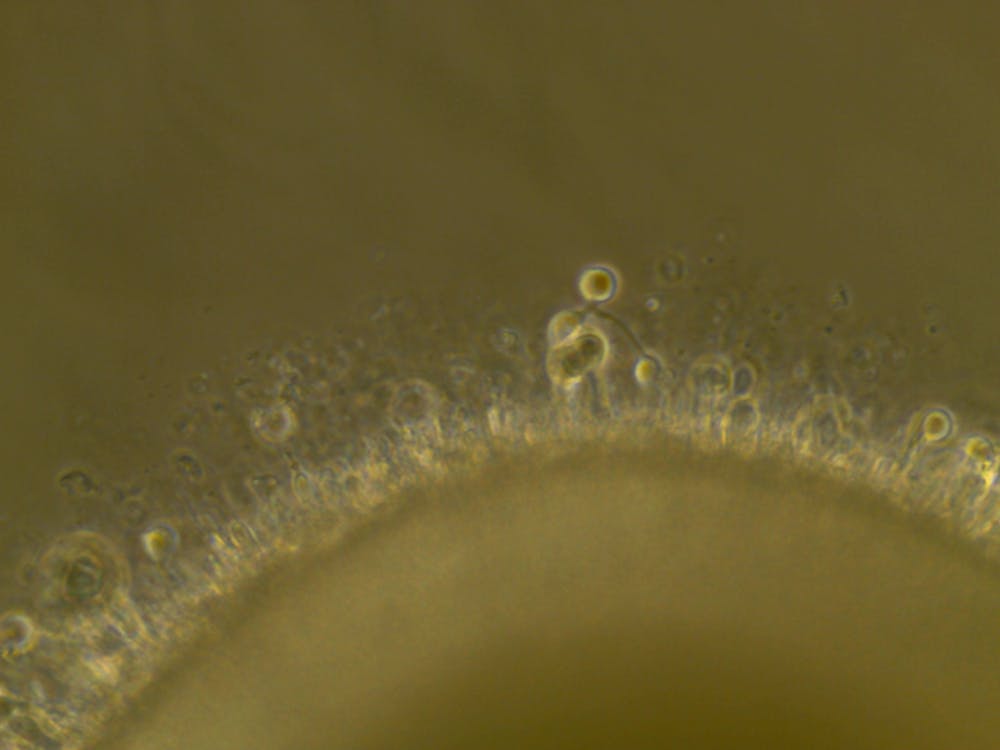
Throughout the process, the lab worked with organoids — engineered tissues grown from stem cells that are capable of replicating the structures of natural tissues. While they may not look like eyes, organoids possess all the cells found in human eyes and develop over the same 9-month time span enabling an accurate model for human development.
“The organoids get a lot of the flash, but really this was awesome work by a great team, and it goes across the different strategies to think about human biology,” Johnston emphasized.
In addition to uncovering the retinoic acid mechanism, the team conducted a survey of red and green cone ratios in a group of 700 adults with normal color vision; these ratios can range from one red for three green to 1:17. Due to the overlapping wavelength ranges of these cells, impact on day-to-day life appears marginal, with sensory systems enabling more variation in gene expression than other biological systems. However, there are differences. For example, the color someone ascribes to tennis balls may be perceived as more yellow (indicative of more red cones) or more green (more green cones).
Looking to the future, Johnston hopes to continue exploring the development of the retina, focusing on the mechanisms that regulate thyroid hormones and retinoic acid. Additionally, he hopes that further organoid research will inform their therapeutic usage.
“We need to know how the cells are made, and two, we need to know if these organoids can make all the cells and what times they make them because it'll have impacts if we want to do transplants,” he said. “The big picture goal for our lab is to figure out as much as we can about how the human retina is made. I just think it's a super cool puzzle, how do you make a hundred different types of something? We're really starting to understand that.”


