On Wednesday, Nov. 29, the In-vivo Cellular and Molecular Imaging Center (ICMIC) hosted a seminar featuring Regents' Professor of Chemistry at Georgia State University Jenny Yang. The talk, titled “Noninvasive Precision Imaging of Microenvironment of Cancer and Metastasis,” discussed biochemical approaches to improving magnetic resonance imaging of cancers.
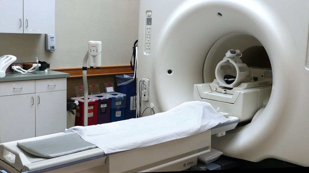
Magnetic resonance imaging (MRI) is a medical imaging technique that uses strong magnetic fields to generate detailed images of soft tissues. In order to improve diagnostic accuracy, radiologists often utilize drugs containing rare metals such as gadolinium called contrast agents to differentiate tissues of interest. According to Yang, current contrast agents have considerable side effects and poor sensitivity to early-stage cancers, driving the need to develop better contrast agents.
“As a protein chemist, the very simple hypothesis that has guided my ambitions is that many key molecular pathways are shared by important biological processes,” Yang said. “I believe that diseases are simple perturbations of these common molecular determinants.”
In other words, imaging the distribution of molecules involved in cancer progression could be a powerful way to diagnose and modulate malignancies in the human body.
Yang’s hypothesis led her to focus on the liver because of its sensitivity to cancers in other organ systems. She explained that many types of cancers metastasize or spread to the liver, which leads to a significant increase in CXCR4 proteins in the liver. This is reflected in an overgrowth of collagen inside and around the organ.
In order to overcome the limitations of molecular contrast agents, Yang turned to proteins. Her team developed ProCA32, a protein containing gadolinium that can be modified to target proteins of interest like CXCR4 and collagen. ProCA32 requires a lower dosage to function and attracts water molecules to enhance its MRI contrast.
“Our contrast agent has really good tumor penetration properties, meaning it can give us additional information about the microenvironment of the cancer,” Yang explained.
When investigating the effectiveness of ProCA32 in human samples, the team found that liver samples stained with ProCA32 targeting collagen showcased greater detail than those stained with Sirius red, the current gold standard in cancer histology. This is exciting because ProCA32 can easily be injected into the body, whereas Sirius red staining requires physicians to extract liver samples for lengthy processing.
These promising results motivated Yang to study a variety of pathological conditions.
Her team has used ProCA32 targeting CXCL12, the ligand to CXCR4, to study the effect of fat on cancer metastasis. Alongside collaborators, Yang’s group imaged the livers of mice fed with low- and high-fat diets with ProCA32-contrast MRI. They found that high-fat mice had a greater number of tumors and more CXCR4 proteins, indicating enhanced metastasis of cancer into the liver. The implications of these results on human health are particularly alarming, given the rapid rise of worldwide obesity.
Yang has expanded this research to the early-stage treatment of liver fibrosis, a major component of most chronic liver diseases. In particular, Yang helped to design a protein drug called ProAgio capable of triggering the death of fibrotic cells. Her team then used ProCA32 to monitor this treatment. The results of this study were extremely fruitful; ProAgio is currently nearing the end of phase I clinical trials.
Yang has also experimented with utilizing protein-based contrast agents for chronic lung disorders.
“Lung imaging largely uses CT… but [this technique] involves heavy radiation and the diagnostic is not very accurate,” she commented.
Most lung disorders involve fibrosis or lung scarring due to excess collagen production. As such, Yang has employed collagen-targeting ProCA32 to accurately visualize lung fibrosis in mouse models of e-cigarette-induced chronic obstructive pulmonary disease and idiopathic pulmonary fibrosis.
Yang closed the seminar by addressing her current and future goals, including introducing her platform technology ProCA to the contrast agents market through her startup InLighta BioSciences. Currently, both ProCA32 targeting collagen and non-targeted ProCA32 have completed the preclinical stages and are in preparation for human trials. Over the past four years, Yang’s group has worked on developing processes to produce, purify and perform quality control on commercially relevant quantities of ProCA32.
“We have a variety of contrast agents built off the same principle, which can have different targeting moiety specificity,” Yang said. “We are really enthusiastic for next year so that we can try for humans to see [ProCA32’s] efficacy.”


