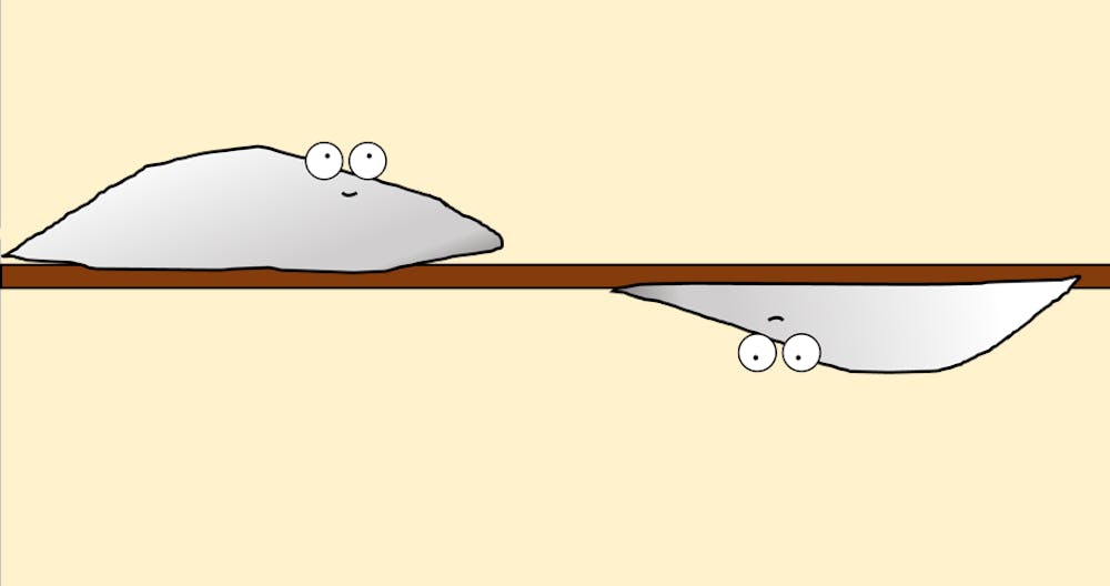
Whether it’s a lab technician staring at a Petri dish from above or a Hopkins student taking notes from a PowerPoint, biology is often only studied from a two-dimensional perspective. A team of scientists at Hopkins and Virginia Tech has begun to shift this perspective with a recent paper exploring cell motility, published in Proceedings of the National Academy of Sciences (PNAS).
The paper examines the long-understood phenomenon of Contact Inhibition of Locomotion (CIL). In CIL, two cells approaching each other will abruptly turn around, almost like repelling magnets.
Brian Camley, a physicist turned biophysicist at the School of Arts and Sciences, had been working with CIL theoretically since 2014. In 2019, he attended a Biophysical Society conference in Baltimore, where he first met Amrinder Nain.
Nain has a mechanical engineering background and currently works in mechanobiology at Virginia Polytechnic Institute. Working with a graduate student, he produced a three-dimensional medium laced with nanofibers to observe cells in. The researchers found that cells traveling on nanofibers did not reverse direction upon contact. Instead, these cells would slide past each other like two busy commuters in a crowded train station.
In an interview with The News-Letter, Nain reported that he and his team were not expecting this outcome.
“I started reading into the literature of contact inhibition,” he said. “And that’s when it hit me that this was really something different that we were observing.”
When Camley saw these data at the 2019 Biophysical Society conference, he was also surprised.
Camley had tried to produce a similar cell response in the past on a flat surface, but he found that Nain’s experiments were much more replicable.
“I found that you really needed to tune behaviors really tightly, and it was a non-generic behavior,” he said. “It was something you had to work really hard to generate. All of the sudden, this happened very naturally.”
After their chance encounter at the conference, Camley and Nain began collaborating remotely, with Camley working on the modeling side of the project and Nain collecting experimental data.
When explaining the determined mechanism of cells scurrying past each other, both Camley and Nain held out both their palms in a synchronized choreography. As the cells were about to collide, they turned on the axis of the nanofiber, where they could both continue in their respective directions. In the PNAS publication, this phenomenon can be most clearly seen in video S28, which can be found in the press release for the study.
Nain remarked at how predictable the phenomenon seems in retrospect.
“Now that we look back, it was so obvious that this would result in this outcome,” he said. “It’s a very simple system, but it has profound outcomes.”
Even with these amazing results, the team acknowledged that there would seldom be only be one fiber in an extracellular environment. When researchers inserted an additional parallel fiber into the matrix, they encountered an entirely different pattern of cell motility. Now, instead of squeezing past each other, the cells stuck together like train cars traveling off in one direction. One simple shift in the environment had produced a profound impact on cell behavior.
Both Camley and Nain acknowledged that their work may explain why some cell studies have different outcomes in the laboratory and in a living organism. For example, in vitro drug studies, also known as “test-tube experiments,” commonly produce different results than in vivo studies that examine the impact of a drug on an intact organism. Although many factors can explain this phenomenon, the behavior of cells in three dimensions as opposed to two may be one explanation why.
According to Nain, he and Camley attempted to recreate a body-like environment within the lab.
“Biology is studied typically on a flat Petri dish, and there are very few instances where you can say a body is a flat 2D,” he said. “So [our objective] is to recover or recapitulate the fibrous environment, and that is a very complex environment that we still don’t know a lot about.”
Following the results of this experiment, the team is working on a grant from the National Science Foundation to examine how strongly cells adhere both to the fibers and to each other. They plan to do this by manipulating the fibers to see how much force it takes to pull cells apart. In addition, Camley expressed an interest in seeing how different chemicals might impact cell motion in this 3D cell playscape.
Camley reflected on the serendipitous nature of the project, which began with a chance encounter.
“The whole reason that this project existed is because of a happenstance of us colliding at a poster session,” he said.
Nain smiled at the word choice. “Us colliding!” he repeated. Much like some of the cells they were studying, the two had decided to stick together after their encounter.


