Social media accounts, such as Johns Hopkins Medicine (JHM) Fundamentals, have introduced a novel approach to drawing larger audiences into the realm of scientific research. Such accounts highlight the images produced in the research process rather than the data collected.
JHM Fundamentals features the latest discoveries in medicine and related biological sciences, supported by the Institute for Basic Biomedical Sciences at Hopkins. In an email to The News-Letter, Vanessa Wasta, the communications and marketing manager, explained the purpose behind the account.
“Our focus primarily on research and science-based content distinguishes us from pre-existing enterprise accounts at the university, setting us apart from other institutions as well,” she wrote.
After almost six months of planning, the team launched the official JHM Fundamentals Instagram account in 2018. The following year, JHM Fundamentals was named the 2019 silver awardee for excellence in social media by Association of American Medical Colleges’ Group on Institutional Advancement Awards for Excellence, a competition honoring academia’s most creative approaches to promote medicine and medical research.
Wasta explained how platforms like Instagram immerse viewers in what she called the “story of science.”
“Through images, we provide a window into the biology that our scientists are investigating in a very different style from narrative content,” she wrote. “Imagery provides an experience in witnessing science.”
Senior Communications Specialist Rachel Butch is the co-founder and primary editor for JHM Fundamentals. She explained that the team chooses images that are aesthetically pleasing and have a compelling discovery story.
“We strategically opt to focus on molecular, cellular and other scientific images rather than people or places,” she wrote in an email to The News-Letter. “We also love images that are visually striking, have a wide range of color and dimension, and those in which you can ‘see’ the biological processes as they occur.”
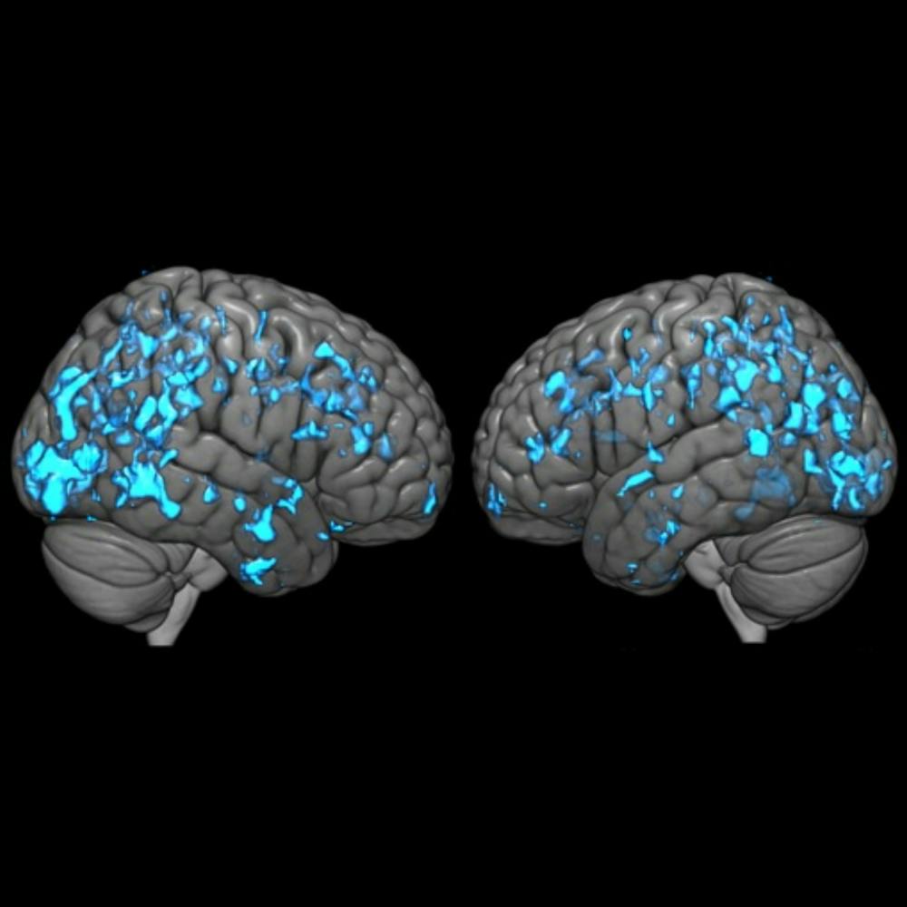
One of the striking images featured on JHM Fundamentals was produced by Richman Family Professor Gwenn Smith of the Psychiatry and Behavioral Sciences Department. In a study published in Parkinsonism & Related Disorders, Smith and her colleagues discerned the effects of deep brain stimulation on metabolism and the levels of dopamine, a neurotransmitter. Deep brain stimulation is regarded as a treatment for Parkinson's disease and some forms of epilepsy.
The researchers created the image using Statistical Parametric Mapping software, which is an image analysis program. The software allows researchers to display statistical results on a brain template.
The blue areas are the brain regions of Parkinson’s disease patients after treatment with deep brain stimulation where an increase in dopamine is associated with an increase in glucose metabolism.
“The unique aspect of the study is that we used 2 PET tracers to highlight different aspects of pathology that are interrelated,” Smith wrote in an email to The News-Letter. “In the imaging field, this illustration would be considered fairly standard but the data that goes into the image is unique.”
PET, which stands for positron emission tomography, is a technique that produces images of processes in the body. From their research the team concluded that Parkinson’s disease patients’ improvement in motor function after deep brain stimulation may be related to the observed increase in metabolism and changes in dopamine levels.
In addition to their posts, the team behind JHM Fundamentals takes advantage of Instagram’s features to ensure that the account’s content reaches its intended audience efficiently. Through tracking data available on Instagram, the team learned that their followers especially liked data visualization images and videos. They use Instagram Stories to highlight scientific happenings at Hopkins, such as the 2019 Nobel Prize win of Gregg Semenza, a professor of Genetic Medicine at the School of Medicine.
The team plans to continue to engage with their audience in new ways, such as creating interactive quizzes and holding a competition to vote for the best images on the platform.
One of the contenders for a potential future competition is a scanning electron micrograph of a mouse trachea, an organ colloquially known as the windpipe. In yellow are goblet cells which produce mucus and in pink are motile cilia. The creation of motile cilia is the focus of study in Andrew Holland’s lab at the School of Medicine.

“This image is a beautiful representative image of the motile cilia that we study in their natural context, the trachea, where they are important for moving pathogens and mucous out of the airway,” Gina LoMastro wrote in an email to The News-Letter.
LoMastro is a PhD student in the Holland lab and one of the co-authors of the paper in which the image was featured. Holland and his team discovered that centrioles — a structure that is required for the production of motile cilia — can be produced in large numbers in the absence of organelles called deuterosomes. The researchers verified their discovery by creating mice genetically engineered without deuterosomes, counting the number of motile cilia in the trachea of those mice and comparing those numbers to the mice with deuterosomes.
“We spend a lot of time perfecting our imaging techniques so they accurately represent what is happening in our experiments and we can make precise conclusions,” LoMastro explained. “Understanding how to perform experiments is key to obtaining high quality images that allow us to both generate rigorous data and beautiful scientific images.”
The process of creating the micrograph was entirely different from the brain image created in the Smith lab — reflecting the diversity of scientific discoveries featured on the JHM Fundamentals account.
To create the micrograph, a sample of the trachea of a mouse was dissected and then fixed. Fixing a biological sample stabilizes its ultrastructure, the minuscule structure within a cell that can only be seen through an electron microscope. Then the sample undergoes a series of dehydration steps and is coated with gold. The gold coating enhances the quality of the image. Once it is fully prepared, the trachea is imaged using the scanning electron microscope. Finally, the resulting image was artificially colored.
“I think biology is fascinating, but as specialists we can get caught up in the details which might not be so easily understood by the public,” LoMastro wrote. “I hope that sharing images demonstrating how beautiful biological processes can be might excite the public and generate enthusiasm for biomedical research.”


