At the intersection of art and science is science imaging, which tells visual stories of scientific processes. However, compared to other art genres, images from science rarely get much attention in art galleries.
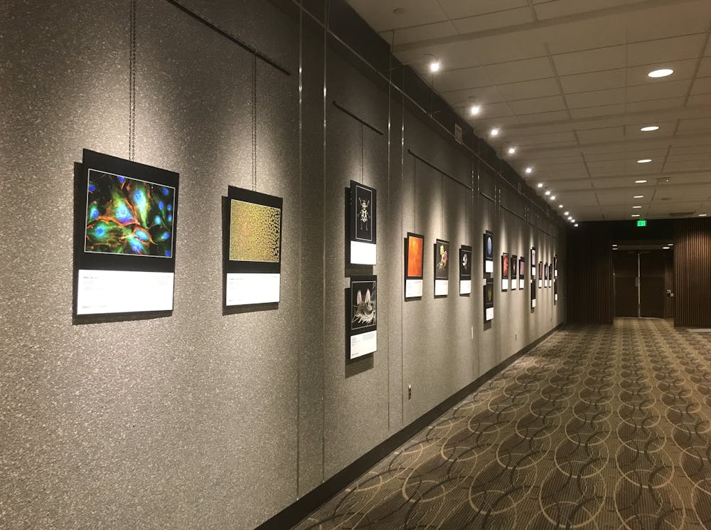
A recent exhibition at Hopkins hopes to increase exposure to the beauty of science images. Until March 20, Images from Science 3 will be holding an exhibit showcasing a variety of scientific images at the School of Medicine’s Turner Concourse.
The exhibit, which opened on Jan. 20, is in collaboration with the Rochester Institute of Technology and Hopkins. It displays scientific images in novel ways in order to engage viewers to learn more about science. It currently takes the forms of an exhibition catalog, a traveling print exhibition and an online gallery.
The first Images from Science exhibition opened on Oct. 12, 2002 in the William Harris Gallery at Rochester Institute of Technology. Professor Michael Peres and Professor Emeritus Andrew Davidhazy founded the traveling exhibit to showcase beautiful, unique images from oceanography, geology, biology, engineering, medicine and physics. During the dawn of digital photography, Images from Science 1 traveled to seven countries.
After the Images from Science 2 exhibit in 2014, the Internet, social media and new imaging technologies rapidly transformed the arena of science imaging.
Images from Science 3 was created through a partnership between Peres and Hopkins professor Norm Barker and built on the successful work of Images from Science 1 and 2. Images from Science 3 is the first in its series to include moving pictures and animations. This year the exhibit is dedicated to Lennart Nilsson, a famous Swedish photographer who was featured in Life magazine.
Scientific imaging from the exhibit includes digital illustrations, confocal and fluorescence photomicrographs, high-speed photographs, astrophotography and a variety of other techniques.
A photomicrograph of bovine pulmonary artery endothelial cells from a cow by Ethan Whitecotton was selected for the exhibit. The image’s vibrancy was achieved through various wavelengths of light hitting the sample. According to the caption for this piece, it seeks to transport the viewer to the cellular level where they learn of the beauty that exists at early, simple stages of life.
A digital illustration poster by Alisa Brandt follows the color changes in Red-capped cardinals from fledging to full adult. The poster was a result of the observation of 25 birds at the National Aquarium in Baltimore. Karl Gaff submitted a photomicrograph of creatine crystals branching and sticking to each other. The crystals formed into a Brownian tree over time and radiated the colors of the rainbow.
The exhibit received over 380 files from 130 contributors from 19 different countries. The submissions came from students, artists, researchers and professors. A panel of Images from Science 3 judges narrowed down the exhibit gallery to 81 images, each judge selecting their favorite image to receive a Juror Selection Award.
The international panel of judges included Hopkins Professor of Pathology and Urology, Jonathan Epstein. The judges evaluated the images online based on aesthetics, uniqueness, degree of difficulty in making and other personal criteria.
Norm Barker, one of the three organizers of the exhibit and Professor of Pathology and Art as Applied Medicine at the Hopkins School of Medicine, explained the importance of the science of imaging in an interview with The News-Letter.
“One could make a compelling argument that imaging has become a science unto itself and is an integral part of the work of every contemporary organization, science image maker and research center,” Barker said.
Barker is a microscopist and has recently published several books and translated them into international traveling exhibits. He finds Stefan Diller’s video animations “Cellular Flybys” to be some of his favorites among this year’s exhibit.
The video animations were created using an electron microscope and combine thousands of still frames of cells.
“It gives one the effect that you are flying over and in and out of cells, which is exactly what you are doing. When you see this on a large screen, it’s awe-inspiring!” he said.
A team of organizers and judges collaborated for 18 months in order to make the exhibit possible. Barker highlighted how planning the exhibit presented challenges to the team.
“The success of the exhibition required constant innovation and problem solving,” he said.
The Image from Science 3 exhibit is making its next stop at the New York Hall of Science before heading to Northern Illinois University’s Jack Olson Gallery. The organizers hope to take the exhibit to Museo Alto Garda in Riva del Garda, Italy.
They are also working on translating the text of the exhibit and printed catalogue into Chinese for potential exhibit displays in Asia. Barker predicts exponential growth in Images from Science exhibits in the future.
“I think the general public is taking a greater interest in science exhibitions in museums as well as science centers. More and more fine art institutions are showing science,” he said.


