Dr. Bernardo Sabatini, a professor of Neurology at Harvard Medical School and a Howard Hughes Medical Institute investigator, gave a talk titled “Basal Ganglia Circuitry for Action Selection and Evaluation” on Oct. 17 at the Hopkins Preclinical Teaching Building at the School of Medicine.
The Sabatini Lab at Harvard studies the basal ganglia and synapses along with the development of imaging and molecular technology to treat neurological diseases. According to his lab website, the mission of his research is to study the structure and function of synapses and how synapse function relates to animal behavior.
Sabatini began the lecture by outlining the scope of his talk, which consisted of the development, function and regulation of basal ganglia. The basal ganglia, rather than a distinct brain structure, instead consists of several groups of “nuclei” or neurons, spread deep within the cerebral hemisphere. According to Sabatini, these structures have two primary functions.
“[They are responsible for] triggering coordinated sequences and motor actions to promote the repetitions of behaviors that lead to reward,” he said.
Unfortunately, the varied and important functions of the basal ganglia also lend way to a number of diseases, including Parkinson’s and Huntington’s. Additionally, reward behavior mechanisms in the basal ganglia are responsible for most forms of addiction. Sabatini explained his belief that autism is the result of basal ganglia processes gone awry.
“Many of the main symptoms… involve repetitive movements and behavioral inflexibility,” Sabatini said.
The basal ganglia connects the cortex of the brain to the corpus striatum. Sabatini also stressed the complexity of the basal ganglia system, involving excitation and inhibition processes from five interconnected regions of the brain.
For most of the talk, he focused on the development of the basal ganglia.
“In the first few years of life, humans tremendously expand their behavioral repertoire and gain the ability to engage in complex learned and reward-driven actions,” Sabatini said.
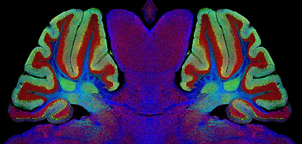
The Sabatini lab uses transgenic mice to study circuitry development, wherein specific regions of their brains are made fluorescent using channel rhodopsins, or proteins involved in the absorption of light. Like humans, mice also develop sophisticated spatial navigation and engage in reward reinforcement learning, although in a much shorter time period.
Brain slices of the mice are taken at four time points, ranging from six to 14 days after birth. By comparing the sum electrical activity of specific regions in the corpus striatum, a key component of the basal ganglia, researchers can understand the rate of neuronal growth and development in the brain.
Still, communication between striatal pathways can go awry. When it comes to diseases such as Parkinson’s and Huntington’s, pathways in the basal ganglia become disconnected, preventing communication between neurons, causing repetitive movements and lack of coordination which are hallmarks of the diseases.
The Sabatini lab used mice with a deletion of the SHANK3 gene, which is a major component of the postsynaptic terminal of glutamatergic synapses in mice. Glutamatergic synapses enable excitatory connections between the cortex and the striatum to perform commands. As a result, Sabatini expected mice without SHANK3 to exhibit reduced levels of neuronal activity. Instead, he explained, the opposite happened.
“During the first 30 days, there was hyperactivation of electrical activity in the SHANK3 mice compared to the mice model,” he said.
Thus, despite the lack of proper communication between neurons, there were more active synapse sites in the SHANK3 mice. However, Sabtini explained, by day 60 this result had reversed.
“There were fewer active synapses than day 30 in the transgenic mice,” he said.
Yet, the normal mice model had continued normal development. Sabatini believes that the reduction of active synapses may be the result of homeostasis of the striatum nuclei with other brain structures, favoring a model in which cells gradually integrate into a circuit.
In that way, synapses remain primarily isolated in brain regions until later development. In SHANK3 synapses, however, the neurons remain continually hyperconnected, potentially causing repetition of movement and actions seen in Parkinson’s and Huntington’s. Despite his theories, Sabatini admitted that his lab had not yet reached a conclusion.
“We don’t know why… yet,” he said.


