Neuroscientists at The Scripps Research Institute (TSRI) recently discovered how serotonin, a brain hormone, can lead to weight loss.
In recent years, biologists have learned that different regions of the brain are able to individually control how your body metabolizes fat. However, until recently, researchers did not understand how the brain was able to do so.
In previous studies, Supriya Srinivasan, a biologist at TSRI, and her team uncovered the 5-HT circuit in the intestine of Caenorhabditis elegans, the signaling pathway for fat metabolism in the roundworm.
In the new study, Srinivasan and her TSRI group identified the required protein and its receptor that stimulate the breakdown of fats in response to serotonin.
Neurons in the brain pick up on certain sensory cues like food availability and produce serotonin in response. Serotonin acts as a messenger, telling a different set of neurons to begin producing the protein, called FLP-7. It circulates through the blood to the intestinal cells and instructs those cells to begin breaking down fat to make energy.
Srinivasan’s team created C. elegans mutants by deleting two genes, unc-13 and unc-31, that are responsible for transporting neurotransmitters and neuropeptides, respectively. In this way, the team could determine whether fat metabolism relies more heavily upon neurotransmitters or neuropeptides.
Results showed that C. elegans with the unc-31 deletion retained almost all of their body fat, indicating that neuropeptides are responsible for stimulating fat metabolism.
From there, the team began to change the expression levels of different genes that code for neuropeptides, looking for the one with the highest fat retention rate. They determined that when the gene flp-7 expresses its corresponding protein, FLP-7, in overabundant amounts, the C. elegans loses most of its body fat.
The FLP-7 protein greatly resembles proteins of the mammalian Tachykinin. About 80 years ago, scientists identified Tachykinin as a protein that caused contractions in the gut. However, no one suspected that it was linked to fat metabolism.
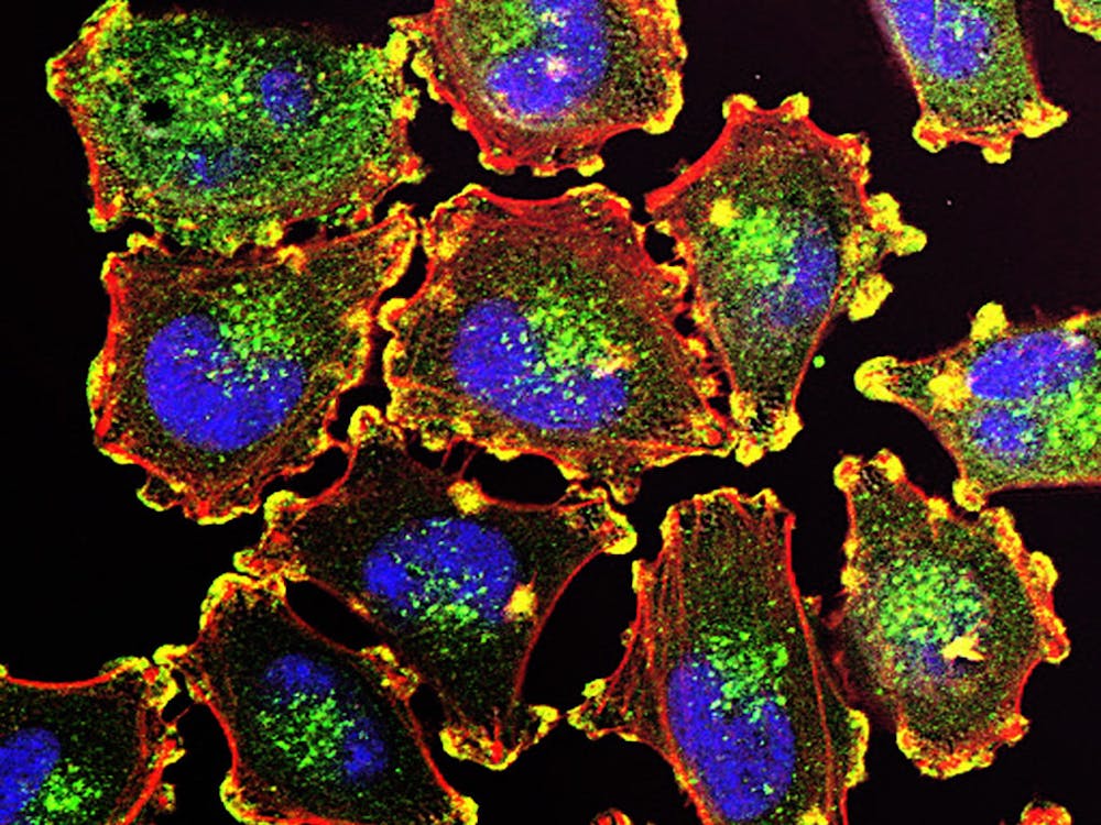
To visualize where in the body flp-7 is active and which neurons are releasing the protein, the team used fluorescent microscopy. They determined that the protein was expressed in neurons in the head but not in neurons in the intestines or muscles.
Next, the team wanted to establish a direct causal link between serotonin levels and FLP-7 expression. Lavinia Palamiuc, a TSRI research associate, used fluorescent microscopy to observe FLP-7 in living animals. Her results suggest that neurons in the brain secrete FLP-7 in response to elevated serotonin levels.
She was then able to visualize how FLP-7 travels through the circulatory system to start the fat burning process in the gut.
Despite mediating fat retention, FLP-7 does not alter food intake, ability to move or reproduction. The research as a whole suggests a novel model for neurons to sense food availability from the environment and subsequently regulate metabolism.
“That was a big moment for us,” Srinivasan said.
The findings in C. elegans could have big impacts on future pharmaceutical production.
Palamuic suggested that a major potential challenge in the future would be to decipher the relationship between nutrient sensing in the nervous system and the mechanisms governing the secretion of each distinct neuropeptide.


Please note All comments are eligible for publication in The News-Letter.